
Figure 7. Observing differential β-gal activity in MCF7-lacZ tumors.
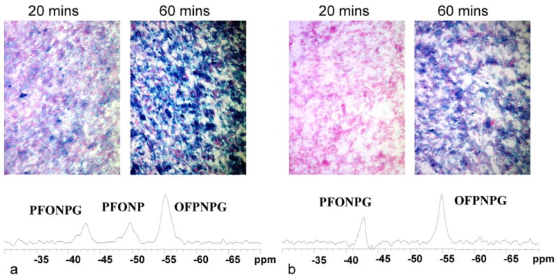
19F NMR spectra are shown for two mice each with a WT- and a lacZ-tumor in contralateral thighs. In each case PFONPG was injected into the lacZ-tumor and OFPNPG into the WT. a) In the first mouse rapid hydrolysis was observed in the lacZ-tumor and β-gal activity was confirmed by intense blue staining with X-gal post mortem (20 and 60 minute development times). b) In the second mouse neither reporter was hydrolyzed indicating lack of β-gal activity in lacZ- or WT-tumor. Histological staining post mortem with X-gal confirmed lower activity, as revealed by the rate and intensity of blue staining.