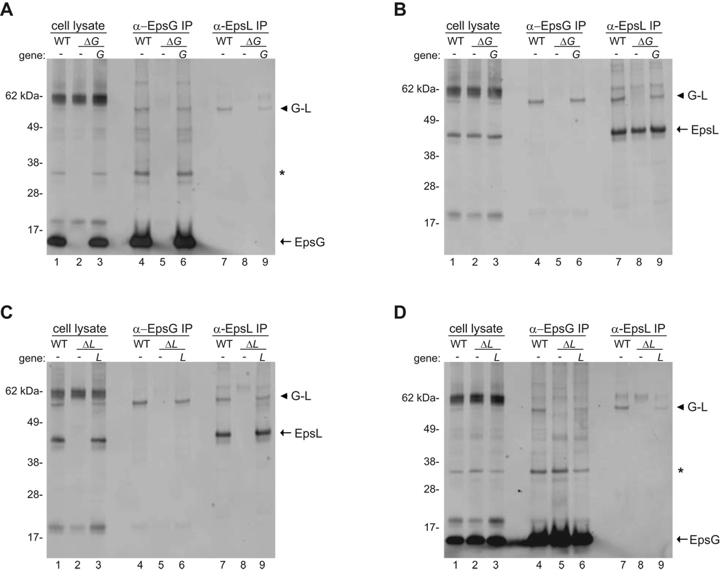
Figure 3. Co-immunoprecipitation of EpsG and EpsL.
Triton X-100 cell extracts were prepared from PBAD::eps wild-type, PBAD::ΔepsG and complemented strain (Panels A and B), and PBAD::ΔepsL and complemented strain (Panels C and D) after cross-linking with 0.25 mM DSP. Cleared cell extracts were immunoprecipitated with either anti-EpsG or anti-EpsL antibodies and subjected to SDS-PAGE and immunoblotting with biotinylated anti-EpsG (Panels A and D) or biotinylated anti-EpsL (Panels B and C) antibodies. In all panels the monomer for EpsG or EpsL is indicated with an arrow and the 60 kDa EpsG-EpsL complex is denoted by an arrow head. In panels immunoblotted for EpsG, the putative EpsG dimer is labeled with an asterisk. The molecular weight markers are shown in kilo Daltons. Lane numbers are indicated.

