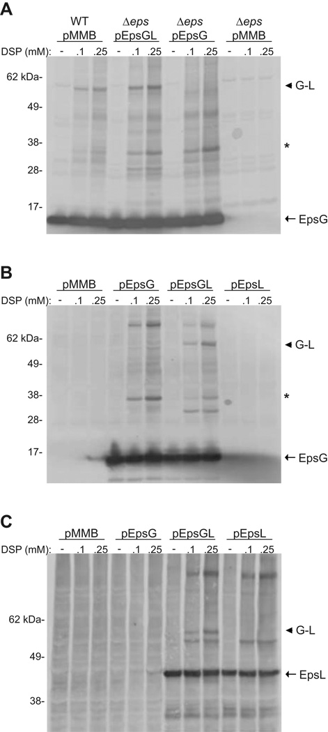
Figure 5. EpsG and EpsL interact in the absence of other T2S components.
(A) Whole cells of V. cholerae TRH7000 wild-type, and TRH7000 with the entire eps operon removed (Δeps) expressing plasmid-encoded epsG, or epsG and epsL (pEpsGL) were cross-linked with the indicated concentrations of DSP and immunoblotted for EpsG. (B and C) Whole cells of E. coli MC1061 with pACYC184-pilD and pMMB67 encoding epsG, epsL, or epsG and epsL concomitantly were cross-linked with the indicated concentrations of DSP and probed with antibodies specific for EpsG (B) or EpsL (C). The monomer for EpsG (Panels A and B) and EpsL (Panel C) is indicated by an arrow. An arrow head designates the EpsG-EpsL complex, and an asterisk represents the putative EpsG dimer in Panels A and B. Positions of molecular weight markers are indicated.

