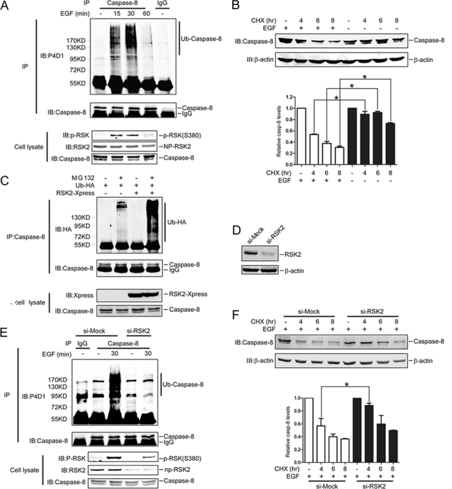
FIGURE 3.
RSK2 is involved in EGF induction of caspase-8 ubiquitination and degradation. A, EGF induces endogenous caspase-8 ubiquitination. HeLa cells pretreated with MG132 (20 μm) for 4 h were immunoprecipitated (IP) under denaturing conditions with anti-caspase-8 after treatment with EGF (100 ng/ml) for different periods of time. Ubiquitination of caspase-8 was detected by Western blot with a P4D1antibody. B, EGF induces caspase-8 degradation. HeLa cells were treated with EGF (100 ng/ml) and CHX (30 μg/ml) for the indicated time, and proteins were extracted. The caspase-8 protein abundance was visualized by Western blot with a casapse-8 antibody. The graph shows data from triplicate experiments (mean ± S.D.). The asterisk (*) indicates a significant difference (p < 0.05, Student's t test). C, RSK2 enhances caspase-8 ubiquitination. HeLa cells were co-transfected with RSK2 and HA-Ub and cultured for 30 h. The cells were treated with MG132 (20 μm) for 4 h, and proteins were extracted. The caspase-8 proteins were immunoprecipitated with a caspase-8 antibody, and ubiquitinated caspase-8 proteins were visualized with anti-HA by Western blot. D, confirmation of RSK2 knockdown. HeLa cells were transfected with si-mock or si-RSK2 and selected in the presence of G418 (800 μg/ml) for 14 days, and then colonies were pooled. The proteins were extracted, and the RSK2 protein level was visualized by Western blot with an RSK2 antibody. Equal protein loading was confirmed by reprobing with a β-actin antibody. E, RSK2 attenuates EGF induction of caspase-8 ubiquitination. Knockdown RSK2 cells that were pretreated with MG132 (20 μm) for 4 h were immunoprecipitated with anti-caspase-8 under denaturing conditions after treatment with EGF (100 ng/ml) for 30 min. Endogenous ubiquitination of caspase-8 was detected with a P4D1antibody. F, RSK2 mediates EGF induction of caspase-8 degradation. Knockdown RSK2 cells were treated with EGF (100 ng/ml) plus CHX (30 μg/ml) for the indicated time, and proteins were extracted. The caspase-8 protein abundance was visualized by Western blot with a casapse-8 antibody. The graph shows data from triplicate experiments (mean ± S.D.). The asterisk (*) indicates a significant difference (p < 0.05, Student's t test). P-RSK, phosphorylated RSK; NP-RSK2, non-phosphorylated RSK2; IB, immunoblot; casp, caspase.

