FIGURE 5.
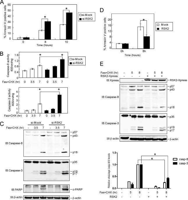
RSK2 blocks Fas-induced apoptosis mediated through caspase-8. A, knockdown of RSK2 induces apoptosis with FasL stimulation. Si-mock and si-RSK2 cells were treated with FasL (500 ng/ml) and CHX (10 μg/ml) and harvested at the indicated time points. Apoptosis was analyzed by annexin V and propidium iodide staining using flow cytometry. Data are represented as means ± S.D. of the percentage of annexin-positive cells as determined in triplicate experiments. The asterisk (*) indicates a significant difference (p < 0.05, Student's t test). B, RSK2 knockdown enhances caspase-8 and caspase-3 activities. HeLa cells stably expressing si-mock or si-RSK2 were treated with FasL (500 ng/ml) and CHX (10 μg/ml) and harvested at the indicated time points. The proteins were extracted, and 50 μg used to determine caspase-8 and -3 activities using the caspase-8 and caspase-3 colorimetric assay kit. Data are shown as means ± S.D. of percentage of activity (1 mg of protein) determined from triplicate experiments. The asterisk (*) indicates a significant difference (p < 0.05, Student's t test). C, RSK2 knockdown enhances FasL-induced cleavage of caspase-8, caspase-3, and poly(ADP-ribose) polymerase (PARP). HeLa cells stably expressing si-mock or si-RSK2 were treated with FasL (500 ng/ml) and CHX (10 μg/ml), and cells were harvested at the indicated time points. The proteins were extracted, and cleaved caspase-8, caspase-3, and poly(ADP-ribose) polymerase (c-PARP) were visualized by Western blot with specific antibodies. β-Actin was used to verify equal protein loading as an internal control. D, overexpression of RSK2 blocks apoptosis induced by FasL treatment. RSK2 was transfected into HeLa cells, and at 30 h after transfection, cells were treated with FasL (500 ng/ml) plus CHX (10 μg/ml) and harvested at the indicated time points. Apoptosis was analyzed by annexin V and propidium iodide staining using flow cytometry. Data are represented as means ± S.D. of the percentage of annexin-positive cells as determined in triplicate experiments. The asterisk (*) indicates a significant difference (p < 0.05, Student's t test). E, overexpression of RSK2 suppresses cleavage of caspase-8 and caspase-3 induced by FasL stimulation. RSK2 was transfected into HeLa cells, and cells were cultured for 30 h. The cells were treated with FasL (500 ng/ml) and CHX (10 μg/ml) for the indicated time, and proteins were then extracted. Cleaved caspase-8 and caspase-3 were visualized by Western blot with specific antibodies. β-Actin was used to verify equal protein loading as an internal control. The graph shows data from triplicate experiments (means ± SD). The asterisk (*) indicates a significant difference (p < 0.05, Student's t test). casp, caspase; IB, immunoblot.

