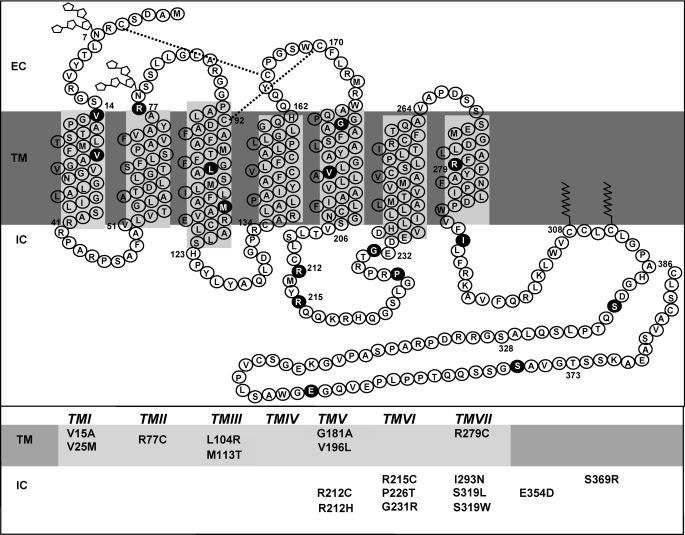
FIGURE 1.
Detected non-synonymous mutations of hIP. Shown is the secondary structure of the human prostacyclin receptor divided into the three major domains: intracellular or cytoplasmic (IC), TM, and extracellular (EC). Glycosylation sites are depicted as ring structures, the dual disulfide bonds are indicated with dashed lines, and the palmitoylation sites in the C-terminal tail are shown as jagged lines. Shown in circles and in the table are the relative positions of the 18 detected non-synonymous mutations analyzed in this study.

