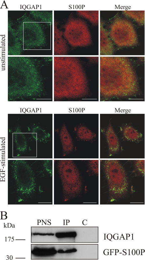
FIGURE 2.
Co-localization and co-immunoprecipitation of S100P and IQGAP1. A, EGF-induced co-localization of endogenous S100P and IQGAP1 to membrane ruffles. HeLa cells were serum-starved for 16 h and either kept unstimulated or were stimulated with 50 ng/ml EGF for 5 min. After fixation and permeabilization, cells were stained with a polyclonal antibody against human IQGAP1 (green channel) and a monoclonal antibody against human S100P (red channel) followed by appropriate fluorescently labeled secondary antibodies. The lower panels show magnifications of the boxed areas. Scale bars, 10 μm. B, co-immunoprecipitation of GFP-S100Pwt with IQGAP1. HeLa cells were transiently transfected with pEGFP-S100Pwt. After 24 h, cells were lysed, and equal amounts of the PNS were subjected to immunoprecipitation (IP) with a polyclonal anti-IQGAP1 antibody or nonspecific anti-mouse IgG polyclonal antibodies (control, C) in the presence of 2.5 μm free Ca2+. The PNS and the precipitated proteins were analyzed by Western blot using a monoclonal anti-IQGAP1 and a monoclonal anti-GFP antibody.

