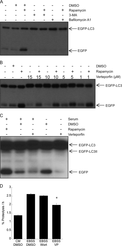
FIGURE 4.
Inhibition of EGFP-LC3 degradation and long lived protein degradation by verteporfin. A–C, MCF-7 EGFP-LC3 cells were treated for 4 h with 30 nm rapamycin without or with 10 mm 3-MA or 100 nm bafilomycin A1 in complete medium (A); different concentrations of verteporfin without or with 30 nm rapamycin in complete medium (B); 10 μm verteporfin in complete medium or in serum-free medium (C). A–C, cells were exposed to 0.1% DMSO as a vehicle control, and EGFP-LC3 processing and degradation were monitored by Western blotting with anti-GFP antibody. D, the amount of [14C]valine-labeled long lived protein degradation was measured in MCF-7 EGFP-LC3 cells treated with 0.1% DMSO, 10 μm verteporfin (VP), or 100 nm wortmannin (Wort) in EBSS, and cells were exposed to complete cell culture medium for 6 h (mean ± S.D. (error bars), n = 3). *, p < 0.05 versus corresponding DMSO treatment.

