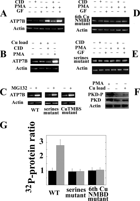
FIGURE 6.
Levels of heterologously expressed ATP7B and endogenous PKD in total lysates of COS-1 cells following experimental variables in vivo. Sixteen hours following delivery of WT ATP7B (A, B, E, and F), 6thCuNMBD mutant (C), Ser-478, Ser-481, Ser-1121, and Ser-1453 mutant (D and E), or CuTMBS mutant (E) cDNA with adenovirus vector, 50 μm CID755673 (CID; PKD inhibitor), 150 nm PMA (PKC activator), 250 nm GF10930X (GF; PKC inhibitor), 20 μm MG132 (proteasome-mediated degradation inhibitor), or 200 μm CuCl2 was added to the culture medium. The cells were harvested 24 h later, and ATP7B expression was evaluated by Western blots of total cell lysates using c-myc tag antibodies (A, B, C, D, and E). Alternatively (F), the levels of endogenous PKD in its phosphorylated (“activated”) form or total PKD protein were evaluated with specific antibodies. Actin expression was also evaluated in the same samples. G, ratios (means and S.D. obtained from four experiments) of ATP7B protein levels in lysates of COS-1 cells expressing WT ATP7B, serine mutant, and sixth copper site NMBD mutant in the absence (−) or presence (+) of 150 nm PMA (PKD activator). Note that PMA increases the level of WT but not the levels of mutants. Error bars represent standard deviations derived from five separate determinations.

