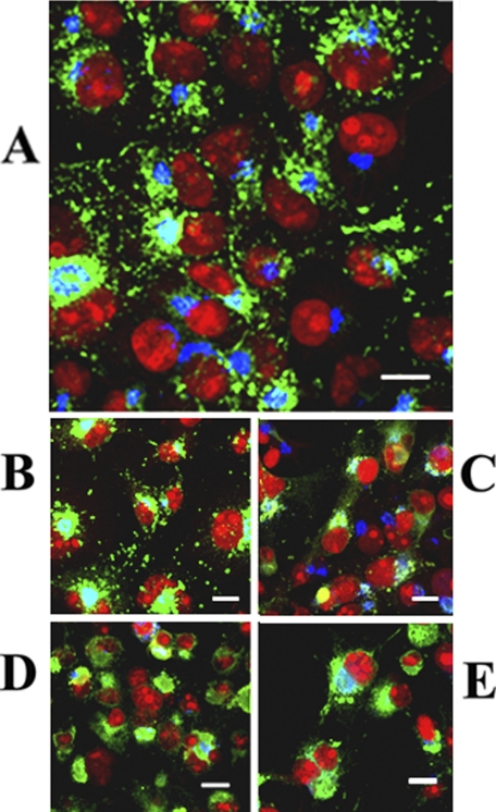
FIGURE 9.
Intracellular distribution of ATP7B in COS-1 cells expressing WT ATP7B (A) or ATP7B subjected to mutations at Asp-1027 (B), at Ser-478, Ser-481, Ser-1121, and Ser-1453 (C), at the transmembrane domain (TMBS) copper site (D), or at the sixth NMBD copper site (E). All panels present different fields of cells treated identically. A copper load (200 μm) was added to the culture media 2 h following infection with adenovirus vector for delivery of ATP7B cDNA (WT or mutant). The cells were fixed 24 h later. Secondary antibodies were goat anti-mouse Alexa Fluor 488 for the ATP7B c-myc tag (green) and donkey anti-rabbit Cy5 for Golgi (blue). Red color indicates nuclei stained with propidium iodide. Scale bar, 10 μm.

