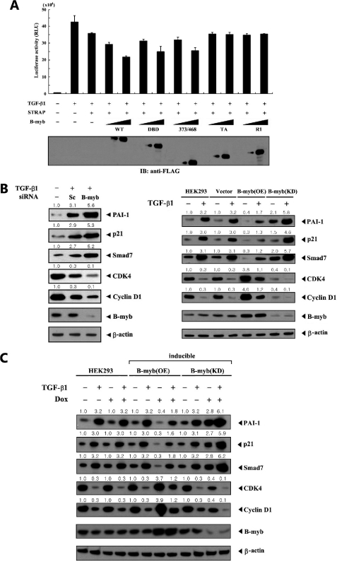
FIGURE 4.
Enhancement of STRAP-induced inhibition of TGF-β signaling by B-MYB. A, HepG2 cells were transiently transfected with 0.3 μg of p3TP-Lux reporter, 0.1 μg of expression plasmid for β-galactosidase as an internal control, and increasing amounts (0.3 and 0.6 μg) of wild-type B-MYB or its deletion mutants (DBD, 373/468, TA, and R1) in the presence of STRAP (0.6 μg), and subsequently stimulated by TGF-β1 (100 pm). Luciferase activity was measured 48 h after transfection and normalized to β-galactosidase activity. The expression level of FLAG-tagged wild-type B-MYB and its deletion mutants was analyzed by anti-FLAG antibody immunoblot. B, effect of B-MYB on the expression of TGF-β target genes. HepG2 cells were transiently transfected with 200 nm of control scrambled siRNA (Sc) or B-MYB-specific siRNA number 1 (B-myb) as indicated, and then treated with 100 pm TGF-β1 for 20 h (left). Cell lysates were subjected to immunoblot analysis using anti-PAI-1, anti-p21, anti-SMAD7, anti-CDK4, anti-cyclin D1, and anti-B-MYB antibodies. Parental HEK293 cells (HEK293), HEK293 cells stably expressing an empty vector (Vector), HEK293 cells stably overexpressing B-MYB (B-myb(OE)), and HEK293 cells stably expressing B-MYB-specific siRNA (B-myb(KD)) were lysed and subjected to immunoblot analysis using the indicated antibodies as described above (right). C, HEK293 cells harboring stably integrated pcDNA4/TO/myc-HisA vector containing wild-type B-MYB (inducible B-myb(OE)) or pSingle-tTS-shRNA vector containing B-MYB-specific shRNA (inducible B-myb(KD)) were cultured in the presence or absence of 1 μg/ml of doxycycline (Dox) for 72 h to determine the effect of B-MYB on the expression of TGF-β target genes. Inducible expression of endogenous B-MYB by doxycycline was assessed by immunoblotting with an anti-B-MYB antibody. Equal amounts of protein in each lane were confirmed by immunoblotting with an anti-β-actin antibody. The relative level of the expression of TGF-β target genes was quantified by densitometric analysis. The fold-increase relative to untreated HepG2 or HEK293 cells was calculated.

