FIGURE 4.
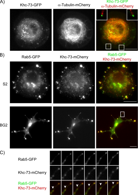
Khc-73 is enriched at the distal ends of microtubules and co-localizes with Rab5-GFP. A, Drosophila S2 cells were transiently transfected with constructs containing mCherry-tagged α-tubulin and GFP-tagged, full-length Khc-73 under the control of the metallothionein promoter (see “Experimental Procedures”). Images shown are an individual frame of a time-lapse movie provided as supplemental Movie 1. Merged panel shows enlargement of boxed regions in the image with Khc-73 puncta at the tips of microtubules. Top left corner corresponds to the box on the lower left, and top right corner corresponds to the boxed region in the lower right. B, Drosophila S2 and BG2 cells were transiently transfected with constructs containing mCherry-tagged Khc-73 under the control of the metallothionein promoter and GFP-tagged Rab5 under the control of the actin promoter. Images shown are individual frames of time-lapse movies provided as supplemental Movies 2 and 3. C, enlargement of boxed region in the merged image of the BG2 cell from B is shown. Arrowhead points to a motile puncta where both Khc-73-mCherry and Rab5-GFP co-localize.

