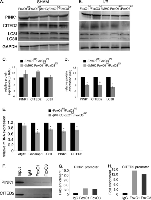
FIGURE 7.
Expression of anti-apoptotic and autophagy-related genes are decreased in βMHC-Cre;FoxO1fl/fl/FoxO3fl/fl compared with FoxO1fl/fl/FoxO3fl/fl mice following I/R injury. A–D, heart lysates were used to examine the protein expression of anti-apoptotic proteins PINK1, CITED2, and an autophagy-related protein LC3II. Immunoblot analyses show a significant decrease in the protein expression of PINK1, CITED2, and LC3II in βMHC-Cre;FoxO1fl/fl/FoxO3fl/fl compared with FoxO1fl/fl/FoxO3fl/fl mice (B) with no significant changes observed after sham procedure (A). C and D are the quantitative representation of A and B, respectively. E, relative gene expression of PINK1, CITED2, LC3II, ATG12, and Gabarapl1 was determined by real time qRT-PCR in βMHC-Cre;FoxO1fl/fl/FoxO3fl/fl and FoxO1fl/fl/FoxO3fl/fl mice. Significance was determined by Student's t test (*, p < 0.05; for FoxO1fl/fl/FoxO3fl/fl, n = 4, and for βMHC-Cre;FoxO1fl/fl/FoxO3fl/fl, n = 6). F, ChIP assay demonstrates that both FoxO1 and FoxO3 bind to the promoter regions of PINK1 and CITED2 under H/R conditions. G and H, graphs are fold enrichment relative to background (IgG antibody control) as determined by qPCR.

