
FIGURE 2.
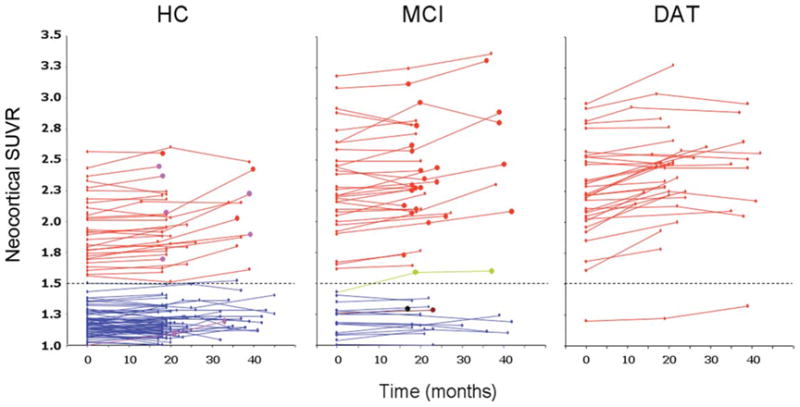
Scatterplots showing individual baseline and follow-up neocortical standardized uptake value ratio (SUVR) values in the healthy control (HC), mild cognitive impairment (MCI), and dementia of the Alzheimer type (DAT) groups. Red full circles in the HC and MCI groups indicate subjects who were reclassified as probable DAT at follow-up. Purple full circles in the HC group indicate subjects who were reclassified as MCI at follow-up. Green, brown, and black full circles in the MCI group indicate subjects who were reclassified at follow-up as probable dementia with Lewy bodies, vascular dementia, or frontotemporal dementia, respectively. Dotted line indicates the SUVR threshold of 1.5 established to separate low [11C]Pittsburgh compound B (PiB) from high PiB participants.