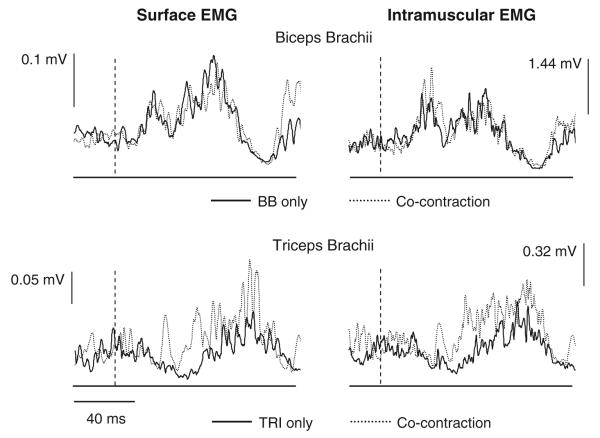
Fig. 4.
Surface (left) and intramuscular (right) electromyographic (EMG) recordings from the biceps brachii (BB) and triceps brachii (TRI) muscles following elbow extension perturbations. The perturbations were all 250 mm/s and 15 mm. The top traces are from the BB muscle; the lower are from the TRI muscle. The intramuscular and surface EMG recordings are the average of the same 20 trials. Responses are shown for both isolated activation of the target muscle (solid lines) and co-contraction of BB and TRI (dotted lines). The dashed lines indicate the onset of the joint perturbation. Note the presence of an excitatory response in surface and intramuscular recordings of the TRI during co-contraction

