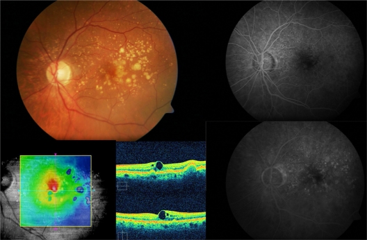
Figure 1.
Subject 3: Baseline visit. A 67-year-old male complained of blurred vision on his left eye. Retinography demonstrates soft confluent drusen within the macular area (top left). Best-corrected visual acuity was 20/50. Optical coherence tomography revealed a drusenoid pigment epithelial detachment, and microcystic hyporreflective spaces within the neurosensory retina and an elevated retinal map (bottom left). Fluorescein angiography did not show any choroidal neovascularization associated (top and bottom right).

