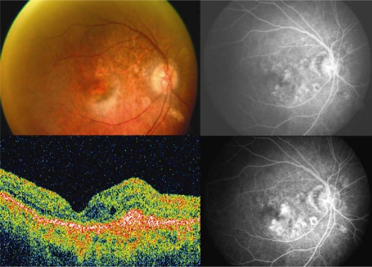
Figure 1.
Case 1: Color fundus photograph of the right eye (upper left) shows a grayish-brown subfoveal lesion suggestive of choroidal neovascular membrane. Early hyperfluoresence with intense late staining on fundus fluorescein angiography confirms the presence of predominantly classic choroidal neovascularization (upper right and lower right). Optical coherence tomography scan reveals the presence of a high reflective, spindle-shaped lesion abutting the fovea with increased overlying retinal thickness and intraretinal cystoid spaces (lower left).

