
Fig. 10.
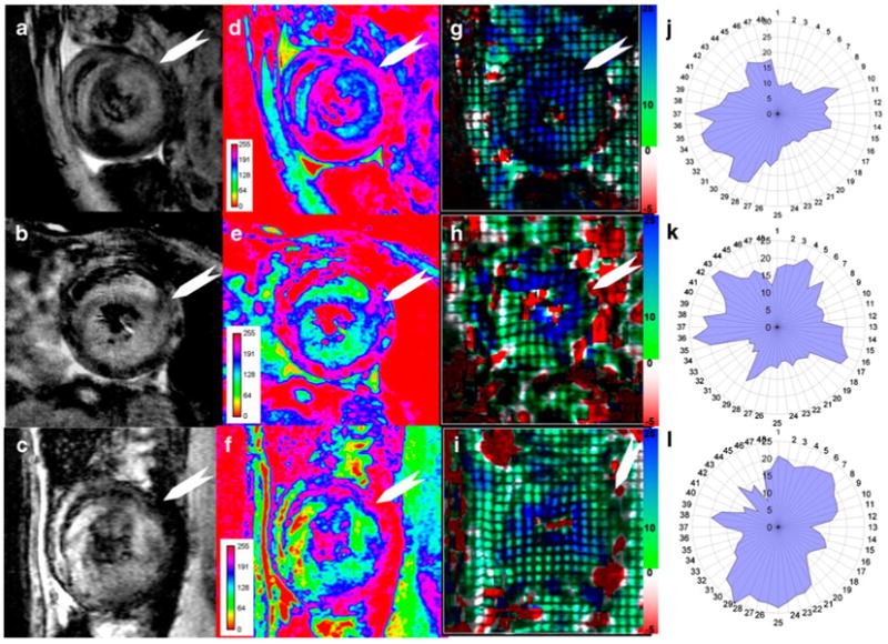
Simultaneous tracking of cellular infiltration and function for 3 allografts: a–c, T2*-weighted MRI; d–f, pseudo-coloring of T2*-weighted images with 8-bit signal-intensity scaling; and g–i, Ecc strain maps. Absolute or positive values of Ecc are indicated: j–l, polar plots of |Ecc|. White arrows indicate the intercepted point of right ventricle and left ventricle, which is the starting location of probe #1. (From Wu et al. [22•]; with permission)