
Fig. 4.
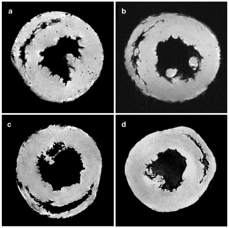
Representative MRM images of transplant and native hearts. Punctate regions of hypointensity can be clearly seen in the allograft heart harvested on POD 109 a after in vivo labeling with MPIO. A representative isograft heart harvested on POD 119 is shown in Panel b. Two native hearts are also shown c and d, one of which was harvested 7 days following MPIO injection c. (From Ye et al. [26•]; with permission)