
Fig. 5.
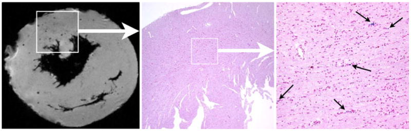
Correlation of MRM and iron staining of MPIO in a POD 94 allograft heart. Image from MRM shows the discrete and circular spots of hypointensity (left panel). These black spots of hypointensity effects are due to the presence of MPIO particles, which was confirmed by the matching section, that correspond to the same area as the boxed region in the MRM image on the left, stained with Perl's Prussian blue for iron (center, ×40 magnification). Right image shows the expansion of the boxed region in center image (×200 magnification). (From Ye et al. [26•]; with permission)