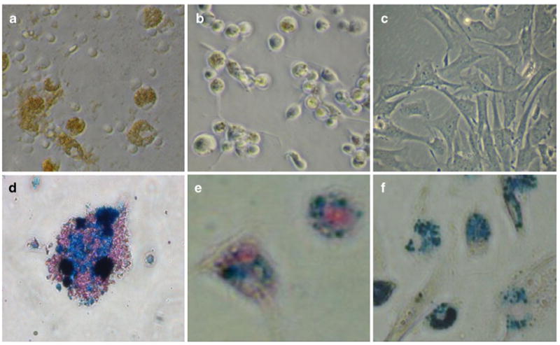
Fig. 8.

Light microscopy of cells labeled with ITRI-IOP. Light microscopy (×200) of a macrophages, b dendritic cells, and c MSCs co-cultured with ITRI-IOP for 24 h, where the brown or light brown color indicates the incorporation of iron-oxide particles inside cells. Light microscope images (×400) of d macrophages, e dendritic cells, and f MSCs after Prussian blue staining also show the presence of iron in the cell cytoplasm (iron shown as blue). (From Chen et al. [48]; with permission)
