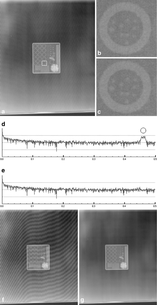
Fig 6.

(a) Mammography Quality Control Phantom (Phantom No. C104, Fuji, Japan) image with grid artifacts. (b) A selected region in (a) is shown in the original resolution. The artifacts are easily seen. (c) The same region, with grid artifacts eliminated. Note that many details, such as the vertical stripes, can be clearly distinguished. (d) The spectrum of a 1-dimensional Fourier transform of the image shown in (a). The y-axis is logarithmic. The frequency of the grid artifact is highlighted with a circle. (e) The spectrum after grid artifacts are removed. (f) An image of the Mammography Quality Control Phantom scaled down 17% to a resolution of 540 × 658 pixels. It shows a very serious artifact (the moiré pattern). (g) The moiré pattern was eliminated using the proposed method. (a) 2,048 × 2,494 pixels; (b) selected area from (a), enlarged. It contains fine vertical stripes. (c) Selected area from (a), with grid artifacts removed. (d) 1-Dimensional Fourier transform of the image in (a). Grid artifact frequency indicated by the circle. (e) The spectrum after grid artifacts are removed.
