
Figure 3.
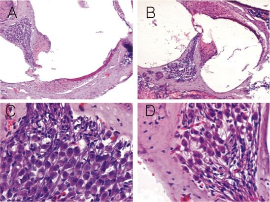
Histology of the cochleae of normal fetuses and fetuses with congenital MCMV infection under HE staining. At low magnification (×100), there are no obvious inflammatory lesions in the cochleae of congenitally infected fetuses (B), but in contrast to normal fetuses (A), the number of spiral ganglion neurons is decreased, and the space among neurons is widened. At high magnification (×400), in contrast to normal fetuses (C), the number of spiral ganglion neurons in the cochleae of congenitally infected fetuses is decreased (D), and the space among them is widened.