
Figure 4.
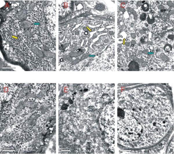
Ultrastructure of the cochlear spiral ganglion neurons of normal fetuses and fetuses with congenital MCMV infection at different ages under electron microscope. Compared with the normal 28-day-old pups (A), swollen endoplasmic reticula (yellow arrow) and increased lysosomes (blue arrow) can be seen in the spiral ganglion neurons of congenitally infected pups (B, C). Compared with the normal 70-day-old pups (D), the spiral ganglion neurons of congenitally infected pups (E, F) show diffused swelling, the physical component of the cytoplasm has disappeared, and a large number of floc and vacuoles has appeared. The number of lysosomes has increased. The chromatins appear condensed and hyperchromatic.