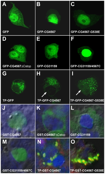
Figure 6. The C-terminal tail of EF-G1 functions as a nuclear localization signal.
(A–I) Subcellular localization of GFP tagged EF-G1 (CG4567) and EF-G2 (CG31159) proteins in living S2 cells. (A) GFP is found in the cytoplasm and the nucleus. (B) GFP-tagged EF-G1 that lacks the N-terminal mitochondrial target sequence predominantly localizes to the nucleus. (C) The mutant CG4567-G538E also mainly localizes to the nucleus. (D) In contrast, when the C-terminal tail of EF-G1 is removed, the GFP-tagged protein is found again in the cytoplasm. (E) Similarly, Drosophila EF-G2 is cytosolic and nuclear. (F) Addition of the CG4567 C-tail to EF-G2 enhances its nuclear localization. (G) Addition of the N-terminal mitochondrial target protein sequence (TP) of EF-G1 results in punctate GFP staining. (H) A similar punctate pattern is seen with a GFP-tagged EF-G1 protein that contains the TP. (I) While punctate staining is also seen with the mutant CG4567-G538E, one can detect a faint signal of the protein in the nucleus (arrow, compare to H). (J–O) Co-localization of GST-tagged EF-G1 and EF-G2 proteins (green) with DAPI (blue, nucleus) and MitoTracker (red, mitochondria) in fixed S2 cells. (J) A GST-tagged EF-G1 protein without the TP exclusively localizes to the nucleus. (K) Removal of the C-tail of EF-G1 changes the subcellular localization from the nucleus to the cytoplasm. (L) Unlike EF-G1, EF-G2 without the TP is found in the cytoplasm. (M) Addition of the EF-G1 C-tail to EF-G2 alters the subcellular localization from the cytoplasm to the nucleus. (N) A GST-tagged EF-G1 protein with the N-terminal TP co-localizes with MitoTracker in mitochondria that aggregate due to fixation (green+red = yellow). (O) The mutant TP-CG4567-G538E also localizes to mitochondrial aggregates (yellow). However, in contrast to the wild type protein, significant amounts of the mutant protein are also detected outside of mitochondria (green).

