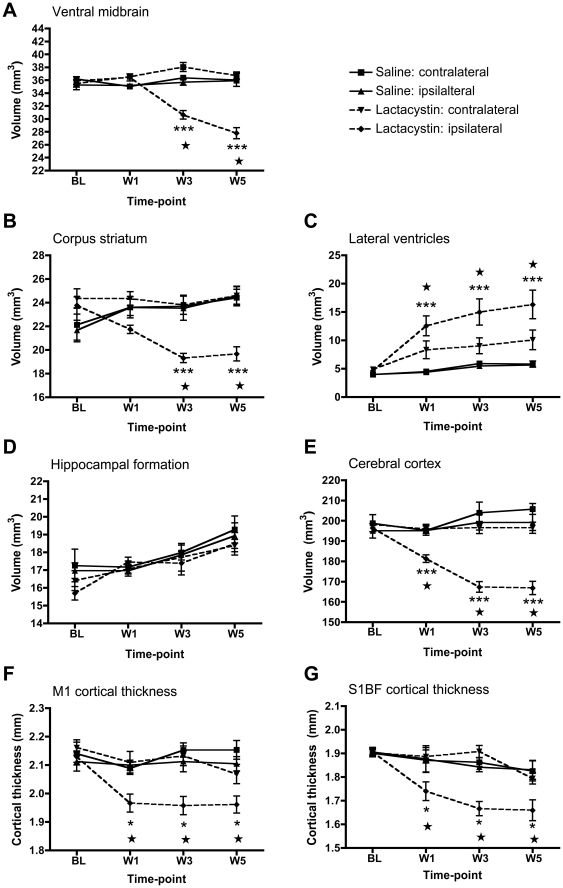
Figure 4. Time-course of regional brain volumetric changes in saline and lactacystin-injected animals.
Significant tissue volume change was observed in (A) the ipsilateral ventral midbrain and (B) ipsilateral corpus striatum at 3 and 5 wks post-surgery compared to the non-injected contralateral hemisphere and both brain hemispheres in saline controls. (C) Lateral ventricle hypertrophy and (D) cortical atrophy were present from 1-wk post-surgery and maintained at 3 and 5 wks in lesioned animals, but not saline controls. (E) No significant change in the volume of the hippocampus was observed in either hemisphere in either group at any time-point. Significant thinning of the primary motor (F) and primary somatosensory cortex were also present from wk 1 post-lesion in lactacystin-injected animals, but not saline controls. Data shown are mean volume (mm3 A–E) or thickness (mm, F, G) ± SEM. ***p<0.001 ipsilateral hemisphere vs. non-injected contralateral hemisphere in lactacystin-injected animals; ★★★p<0.001 ipsilateral hemisphere of lactacystin-lesioned animals vs. ipsilateral hemisphere of saline controls. Saline, N = 5, lactacystin, N = 7.

