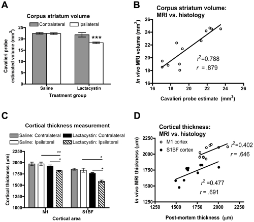
Figure 6. Post-mortem confirmation of in vivo MRI signal changes.
(A) Cavalieri estimator probe measurement of corpus striatum volume post-mortem in saline and lactacystin-injected animals reveals a significant decrease in the volume of the ipsilateral striatum in lesioned animals, (***p<0.01). (B) Linear regression analysis reveals a strong correlation between measurement of striatal volume in both groups from either MRI or post-mortem histology (r = . 879). (C) Cortical thickness measurements post-mortem confirms cortical thinning in the M1 and S1BF cortices of lactacystin-lesioned animals but not saline controls, consistent with MRI data. (*p<0.05; **p<0.01). (D) Linear regression also reveals a robust correlation between cortical thickness measurements in the M1 and S1BF by MRI or from post-mortem histological sections (r = . 646 and .691; respectively). Data shown in (A) and (C) are mean volume or thickness, respectively, ± SEM. Saline, N = 5, lactacystin, N = 7.

