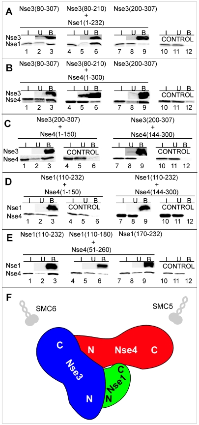
Figure 1. Interactions between Nse1, Nse3 and Nse4.
The indicated His-S-tagged fragments of Nse3 (A, B, C) or Nse1 (D, E) were bound to S-protein agarose-beads and then incubated with in vitro translated Nse1 (A) or Nse4 (B–E). The reaction mixtures were analysed by SDS–12% PAGE gel electrophoresis. The amount of His-S-tagged protein was analysed by immunoblotting with anti-His antibody and the in vitro translated proteins were measured by autoradiography. I, input (5% of total); U, unbound (5%); B, bound (40%). Control, no His-S-tagged protein present. (F) Cartoon of interactions based on panels A–E and our previous work [13].

