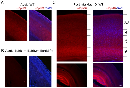
Figure 1. EphB2 is expressed in the developing and adult mouse cortex.
A. Anti-EphB2 immunostaining of a sagittal brain section from adult mouse cortex. B. Anti-EphB2 immunostaining of a sagittal brain section from adult mouse cortex in an animal lacking EphB1-3. C. Immunostaining of a coronal brain section from P10 mouse cortex. Panels on left show EphB2 immunoreactivity in red. Right panels are merged images of EphB2 immunoreactivity and DAPI labeled nuclei in blue. In C, bottom panels show a lower magnification view of EphB2 immunostaining in the cortex and hippocampus of P10 animals. Scale bars = 100 µm in panels A, B, and upper C. Lower C scale bar = 200 µm.

