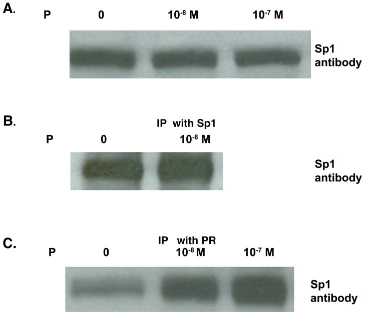
Fig. 6.
Interaction of SP1 protein with progesterone receptor.
T47D cells were plated and grown for 24 hr in media containing 5% FBS. Media was changed to 1% CSS, then to 0.1% CSS, and followed by treatment with either ethanol vehicle or 10−8 or 10−7 M P for 1 hr.
A. Nuclear extracts were subjected to gel electrophoresis and western blot analysis performed using antibody against Sp1.
B. T47D cells were grown as in (A) with final incubation with either ethanol vehicle or 10−8 M P for 1 hr. Nuclear extracts from both treatments were incubated with Sp1 antibody conjugated to Protein A agarose beads. Bound proteins were released by boiling and subjected to western blot analysis probed with the Sp1 antibody.
C. Nuclear extracts from cells treated with either ethanol vehicle or 10−8 or 10−7 M P for 1 hr were incubated with PR antibody conjugated to Protein A agarose beads. Bound proteins were released by boiling and subjected to western blot analysis probed with the Sp1 antibody.

