Figure 2.
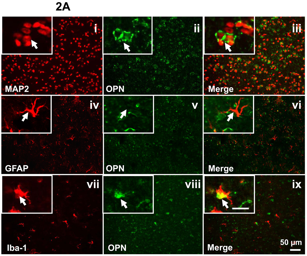
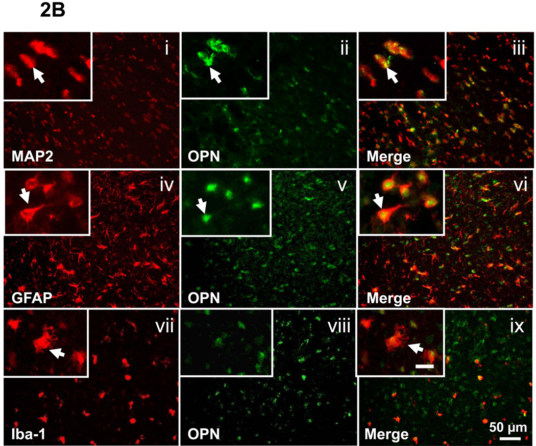
Endogenous OPN expression in P7 sham-operated group (A) and at 48h following HI (B). Double fluorescent labeling for OPN with MAP2 (i-iii), GFAP (iv-vi) and Iba-1 (vii-ix) in the cortex of the ipsilateral hemisphere is shown (scale bar represents 50 µm). In the brain of sham-operated P7 rats, OPN expression was co-localized with the neurons and macrophage (2A, iii and ix, as indicated by arrowhead), but not with the astrocytes (2A, vi, as indicated by arrowhead). At 48h after HI, OPN expression is co-localized with neurons, astrocytes, and macrophage (2B, iii, vi, and ix, as indicated by arrowhead). Insets in the left corner of i-ix are higher magnification (scale bar represents 20µm).


