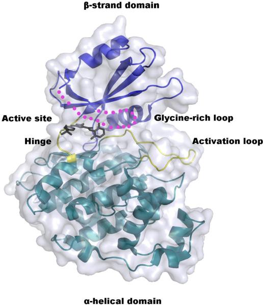
Fig. 4.
Crystal structure of LmajGSK-3 short modeled with the CDK1/2 Inhibitor III. The α-helical and β-strand domains are colored cyan and blue, respectively. The hinge and activation loop of the active site are highlighted in yellow. The glycine-rich loop that is disordered in the crystal structure is depicted as magenta dots. The CDK1/2 Inhibitor III is absent from the crystal structure, but drawn here to depict the location of the inhibitor-binding site.

