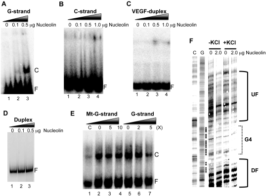
Figure 4.
(A) Nucleolin binding to the intramolecular G-quadruplex structure formed by the G-rich strand of the VEGF proximal promoter region. The VEGF G-quadruplex incubated with various concentrations of nucleolin to determine the formation of the complex with nucleolin. (B–D) The complementary VEGF C-rich strand (B), VEGF oligomer duplex (C) and PCR-amplified duplex DNA representing the proximal promoter region of the VEGF gene (D) were incubated with various concentrations of nucleolin to determine the formation of complex with nucleolin. (E) Competition experiments to measure the specificity of nucleolin binding to the VEGF G-quadruplex. Two different amounts of competitors were added to the binding reaction with 1 μg nucleolin. Lanes 2–4 are competition experiments with mutant G-strand (Mt-G-strand) competitors at 0×, 5× and 10×, respectively, and lanes 5–7 are competition experiments with the VEGF-G-strand at 0×, 2× and 5×, respectively. The ‘C’ and ‘F’ in panels A–E represent the complex and free form of DNA with nucleolin, respectively. (F) Nucleolin binding to the pPu/pPy tract of the VEGF proximal promoter region in supercoiled plasmid DNA. DNase I footprinting was used to determine the binding of nucleolin to the promoter region of the VEGF gene with the plasmid pGL3-VEGFP, which contains the VEGF promoter region from –787 to +50. The bracket ‘G4’ represents a proposed G-quadruplex-forming region and brackets ‘UF’ and ‘DF’ represent up- and downstream flanking regions of the G-quadruplex-forming sequence. Data shown are representative of at least two experiments.

