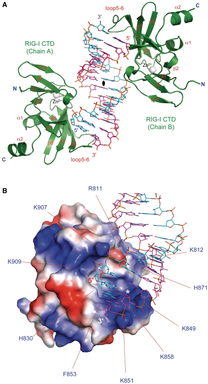
Figure 2.
Crystal structure of human RIG-I CTD bound to a 14-bp blunt-ended dsRNA. (A) Structure of RIG-I CTD bound to the dsRNA. RIG-I CTDs are shown by the green ribbons. The dsRNA is shown by the sticks representation. Carbon atoms of the two RNA strands are colored cyan and pink, respectively. The zinc ion bound to RIG-I CTD is shown by the gray sphere. The complex exhibits pseudo 2-fold non-crystallographic symmetry. The pseudo 2-fold axis is shown by the black oval. (B) Surface electrostatics of RIG-I CTD. Positively charged surface is colored blue and negatively charged surface red. The blunt-ended dsRNA bound to RIG-I CTD is shown by the stick models. Key residues mediating blunt-ended dsRNA recognition are labeled. Location of the ppp-binding site for 5′ ppp dsRNA is indicated by the white asterisk.

