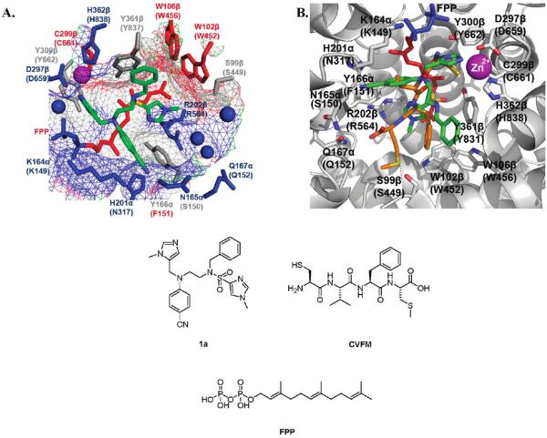
Figure 1.
One high scoring active site conformation of inhibitor 1a (green) as identified by flexible ligand GOLD32 docking experiments (A) using a Connoly analytical surface graphical representation developed in PyMOL,37 red hydrophobic to blue hydrophilic, and (B) using a “cartoon” graphical representation developed in PyMOL37 and overlaid with the peptide inhibitor CVFM (orange) from the rFTase crystal structure. Binding surface of rat FTase (PDB ID: 1JCR35), values in parentheses refer to corresponding residues in PfFTase;31 small molecules colored by atom type: FPP colored red (farnesyl) and blue (pyrophosphate); blue spheres = water molecules; purple sphere = zinc ion.

