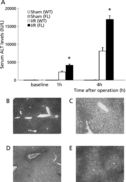
Fig. 4.
(A) Hepatocellular injury as indicated by serum levels of alanine aminotransferase (ALT). Samples were obtained from mice undergoing the sham operation or ischemia for 90 min and reperfusion (I/R). Values represent the mean ± SEM with n = 6 per group. *p<0.05, compared to the WT group. Histological findings of the liver assessed by hematoxylin and eosin staining. (B) Sham operated WT group. (C) Sham operated FL group. (D) WT group 4 h after reperfusion. (E) FL group 4 h after reperfusion. Original magnification ×100.

