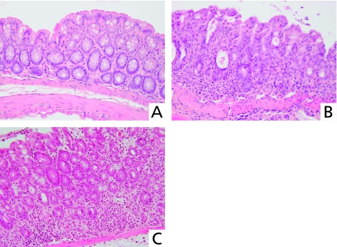
Fig. 3.
Histological findings in the colon of DDS mice. Normal colon mucosa of untreated mouse (A). Colon specimen from DSS mouse shows diffuse crypt disappearance and inflammatory cell infiltration (B) indicating severe colitis. Colon specimen from mouse given oral DSS and daily rectal rebamipide, shows minimal crypt disappearance and inflammatory cell infiltration (C).

