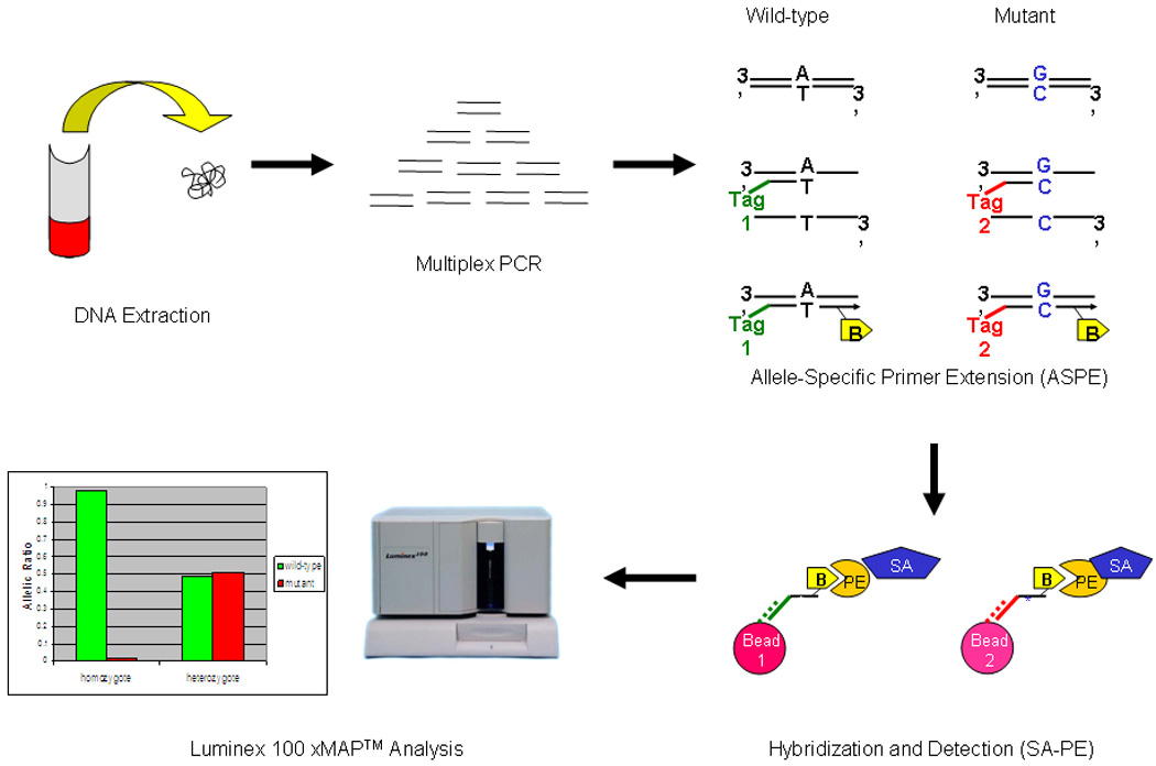
Figure 1. The Bead-based Assay.

Regions of interest are initially amplified by multiplex PCR (for CDKN2A, the primers are listed in Table S1). Individual probes with the interrogative terminal codon are linked to a unique 24-mer Tag and subjected to ASPE. The ASPE extension step uses biotinylated dCTPs thereby introducing a biotin-labeled moiety into the extended fragment. The 24-mer Tag/biotinylated ASPE fragment is then specifically captured by a color-coded bead adorned with an anti-Tag which fully complements the 24-mer Tag sequence. Streptavidin-conjugated phycoerythrin (SA-PE) is then introduced to detect an intensity signal. In summary, the bead color defines the address (i.e. the individual SNP) while the PE fluorescence intensity defines the amount of the specific SNP fragment.
