Figure 1.
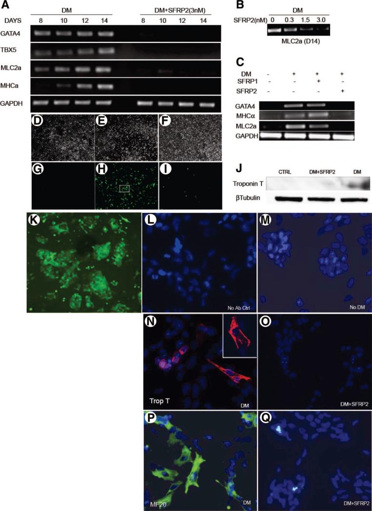
SFRP2 inhibits myogenic differentiation of P19Cl6 cells. Representative reverse transcription-polymerase chain reaction (n = 3) demonstrating (A): expression of myogenic transcription factors and structural proteins in P19Cl6 cells treated with differentiation medium (DM) in the absence or presence of SFRP2 (3 nM), (B): expression of MLC2a in P19Cl6 cells treated with DM for 14 days in the absence or presence of increasing concentrations of SFRP2, and (C) expression of myocyte-specific structural proteins and transcription factors in P19Cl6 cells treated with DM for 14 days in the presence of SFRP1 (3 nM) or SFRP2 (3 nM). (D–I): Phase-contrast views (×100) and green fluorescent protein (GFP) expression in MLC2v-GFP P19Cl6 cells at (D, G) baseline; (E, H) treated with DM; (F, I) DM and SFRP2 (3 nM) for 14 days (n = 3). (J): Western blotting for cardiac Troponin T expression in P19Cl6 cells treated with DM ± SFRP2; CTRL represents untreated cells. (K): High-power view (×200) of (H) inset showing GFP-positive MLV 2v cells. (L–Q): Immunofluorescent staining for Troponin T (red) and MF20 (green) in P19Cl6 cells treated with DM for 14 days. (L): Cells stained without adding primary antibody. Cardiac Troponin T staining of cells: (M): grown in growth medium without induction of differentiation, (N): treated with DM (N inset) zoomed image of Troponin T-positive cardiomyocyte, (O) treated with DM + SFRP2 (3 nM). MF20 staining of cells; (P) treated with DM; (Q) treated with DM + SFRP2 (3 nM). (Nuclei are stained with DAPI for L–Q; total magnification of ×400 for fluorescent images). Abbreviations: Ab, antibody; CTRL, control (untreated cells); DM, differentiation medium; GAPDH, glyceraldehyde-3-phosphate dehydrogenase; MHCα, myosin heavy chain; MLC2a, myosin light chain; Trop T, troponin T.

