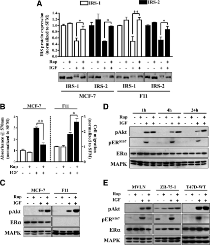
Fig. 1.
Rapamycin blocks IGF-induced proliferation and ERα phosphorylation. A, MCF-7 and MDA-231BO (F11) cells were serum starved overnight and pretreated with rapamycin (Rap; 10 nm) for 30 min before IGF stimulation (4 h). Lysates were collected and resolved by SDS-PAGE for immunoblot analysis against the proteins of interest. The graph represents IRS protein levels as fold change of treated vs. nontreated control. B, Monolayer proliferation was measured by MTT assay in MCF-7 cells (left axis), and motility was determined by transwell Boyden chamber in F11 cells (right axis) in response to rapamycin and IGF treatment. The graph is presented as fold change response vs. nontreated control. C, After rapamycin pretreatment, MCF-7 (left panel) and F11 (right panel) cells were exposed to IGF (1 h) and immunoblot analysis performed. D, MCF-7 cells were serum starved overnight, pretreated with rapamycin for 30 min, and treated with IGF for the indicated time points. Lysates were resolved by SDS-PAGE and immunoblotted against the proteins of interest. E, Western blot analysis of MVLN, ZR-75-1, and T47D cells exposed to IGF (1 h) was performed after 30 min of rapamycin pretreatment. Error bars represent sd on all graphs. The MAPK (anti-MAPK) served as a loading control for immunoblot experiments, and all results are representative of at least three independent replicates.

