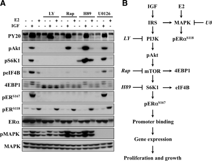
Fig. 7.
IGF phosphorylates ERα via the mTOR/S6K1 axis. A, MCF-7 cells were cultured under hormone-free conditions for 3 d before overnight serum starvation. Cells were pretreated with LY294002 (LY; 10 μm), rapamycin (Rap; 10 nm), H89 (10 μm), or U0126 (10 μm) for 30 min before IGF or E2 (1 h) exposure and subjected to immunoblot analysis. MAPK (anti-MAPK) served as a loading control for all experiments, and results are representative of at least three independent replicates. B, Model of proposed pathway.

