
Figure 2.
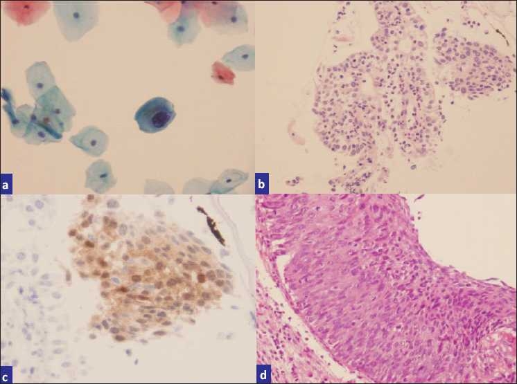
a) Pap smear interpreted as LSIL, b) H and E cell block sections, c) p16-stained cell block sections, d) biopsy showing CIN II-III.
Official websites use .gov
A
.gov website belongs to an official
government organization in the United States.
Secure .gov websites use HTTPS
A lock (
) or https:// means you've safely
connected to the .gov website. Share sensitive
information only on official, secure websites.
a) Pap smear interpreted as LSIL, b) H and E cell block sections, c) p16-stained cell block sections, d) biopsy showing CIN II-III.