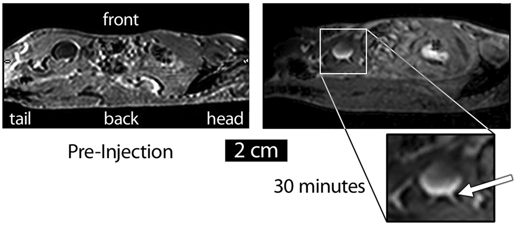
Figure 3.
Sagittal MR image of a C57BL/6 mouse injected IP with APMS-DTPA/Gd-TEG. The particles appear as a bowl-shaped region of contrast in the bladder, and the ureter is also visible (white arrow). Because the animal was lying on its back, these images suggest that the particles were settling under the influence of gravity.

