
Fig. 1. NTPDase1 is the major ectonucleotidase on mouse peritoneal macrophages elicited with thioglycollate.
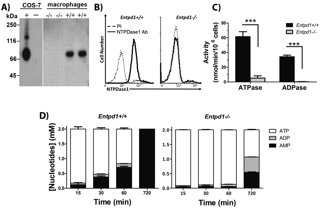
A) Western blot with rabbit polyclonal Ab against mouse NTPDase1 (mN1-2c). Left gel shows control proteins (0.5 µg) from lysates of COS-7 cells transfected with mouse NTPDase1 (+) or untransfected (−). Right gel shows a representative western blot (out of three performed with cells from individual mice) of proteins (12.5 µg) from peritoneal macrophage lysates.
B) Flow cytometric anaylsis of NTPDase 1 analyis using non permeabilizing conditions with a polyclonal Ab against CD39 (C9F, ——), compared to its pre-immune serum (PI, ----). Data are representative of three independent experiments with pooled macrophages from 2 to 4 mice per experiment. C) NTPDase1 activity with either ATP or ADP as substrate, Entpd1+/+ (filled bars) and Entpd1−/− (grey bars) macrophages. Data show mean + SEM (n=3). ***p<0.001, two-way ANOVA with Bonferroni post hoc test. D) Time course of the hydrolysis of 2 mM ATP by Entpd1+/+ or Entpd1−/− peritoneal macrophages was followed for 12 hours by HPLC: ATP (□), ADP (■), AMP (■). No adenosine production was detected. Data show mean + SEM of three independent experiments.