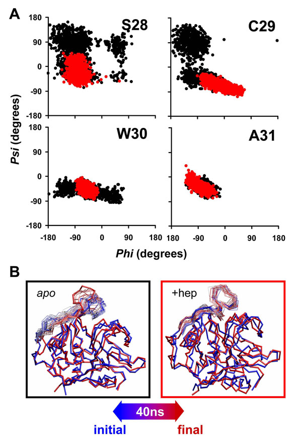
Figure 7.
Heparin binding stabilizes helical content in the active site and occluding loop A) Phi-Psi distributions for the first residues of the main alpha helix (residues 28 to 45) in simulations with protonation corresponding to alkaline conditions. In black : apo catB; In red catB + heparin (R-domain). We present here the only the first four residues of the helix (S28, C29, W30 and A31) since the analysis on the subsequent residues did not shown observable differences in the distributions. B) Superposition of structural snapshots collected during MD, highlighting motions of the occluding loop. The color code from red to blue reflects the time evolution of the trajectory.

