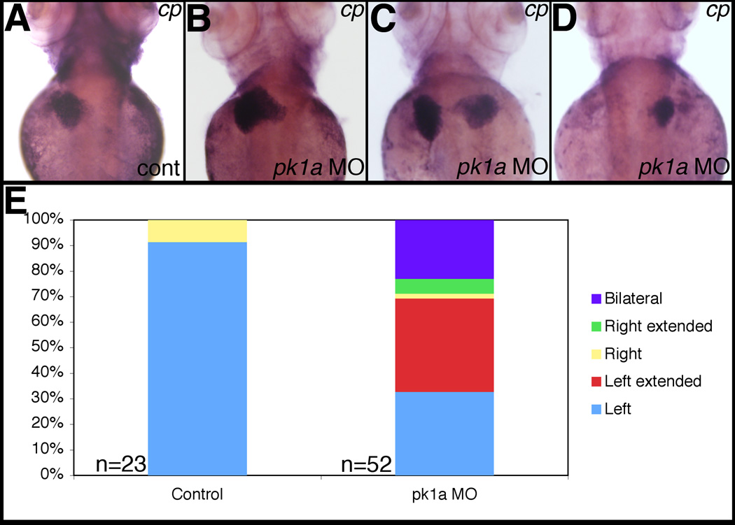
Figure 3. Abnormal liver localization in pk1a morphants.
Whole-mount in situ hybridization of ceruloplasmin (cp), a liver marker, in 3 dpf pk1a morphants demonstrates abnormal liver location, including “right extended” (B), “bilateral” (C), and “right” (D). (E) Graph depicting the scoring of pk1a morphants demonstrates a significant difference in the total number of abnormally localized livers (9% vs. 67%, p<0.0001 by chi-square test), while the difference in livers limited to the right side is not significant (9% vs. 8%). Please see supplemental data for localization defects in other organs.

