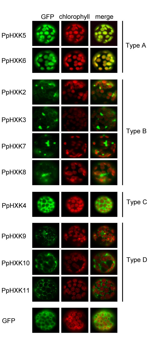
Figure 4.
Intracellular localization of Physcomitrella hexokinase-GFP fusions. Fluorescence microscopy pictures of wild type protoplasts transiently expressing different GFP fusions. GFP fluorescence is shown in green, with the chlorophyll auto-fluorescence in red as a chloroplast marker. Protoplasts expressing GFP alone were also included as a control. The white bars represent 5 μm.

