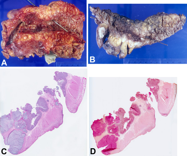
Figure 2.
Segment of colon. (A) Gross, with probe through site of perforation and obstructing ulcerated mass to the left of (distal to) the perforation, (B) Gross, with longitudinally sectioned colon showing relationship between the perforation (with probe) on the right and the obstructing ulcerated mass on the left, (C) Microscopic, with the proximal perforation on the right and the distal transmurally invasive adenocarcinoma on the left (H&E, 1×), (D) Microscopic, same section as (C), showing the mucinous nature of the carcinoma (mucicarmine, 1×) 14.

