
Figure 2. Immunohistochemically stained sections of centrifuged (indicated as C) and uncentrifuged (indicated as U) aggregates.
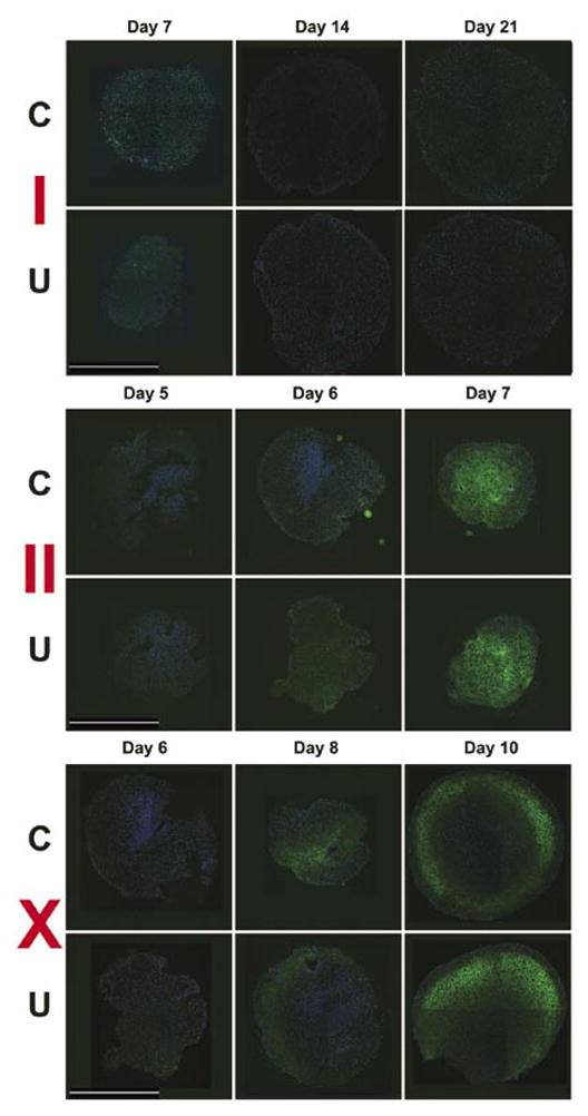
Immunoreactivity for collagen types I, II, and X was detected using a fluorescein isothiocyanate (FITC)-labeled secondary (green) antibody; 4′,6-diamidino-2-phenyl-indole (DAPI) counter-stain of the nuclei (blue). Scale bar, 1 mm.