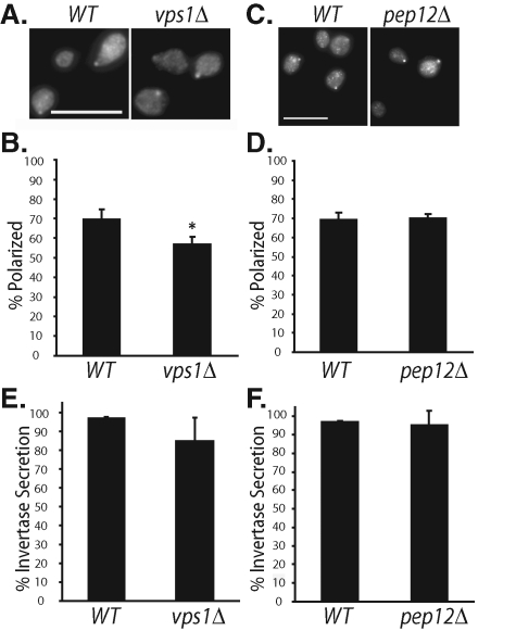
FIGURE 3:
Polarized localization of Cdc42p in vps1Δ and pep12Δ cells. (A) The WT and vps1Δ cells were grown to log phase, fixed, and immunostained for Cdc42p. (B) Quantification of Cdc42p polarization in WT and vps1Δ cells. Fifty small-budded cells were counted for each group (n = 3). Error bars represent standard errors. *, p < 0.05. (C) The WT and pep12Δ cells were immunostained for Cdc42p. (D) Quantification of Cdc42p polarization in WT and pep12Δ cells. Fifty small-budded cells were counted for each group (n = 3). (E) Percentage of invertase secretion in WT and vps1Δ cells (n = 3). (F) Percentage of invertase secretion in WT and pep12Δ cells (n = 3). Error bars represent standard errors.

